A wound can shrink substantially without closing completely. A treatment can improve a wound-area measurement without establishing how many people achieved durable healing. Those distinctions matter when reading research on GHK-Cu, a copper–peptide complex whose clinical and laboratory evidence spans very different settings. The central question is not whether one impressive percentage sounds persuasive, but what was measured, in whom, against which comparison, and at what point in follow-up.
The diabetic-ulcer evidence offers a useful case study in those questions. This analysis uses the indexed primary report abstract and an author-posted full-text transcription, alongside indexed abstracts for a laser-resurfacing trial and two cell-culture studies. The ulcer transcription adds important design information, but its imperfect optical character recognition prevents confident use of some tables. The resulting evidence supports a narrowly framed clinical signal—not an ingredient-wide verdict on facial products, other formulations, or systemic injections.
Study facts at a glance
- Publication year: The ulcer paper was published in 1994; its PubMed identifier does not make it a 2006 trial. [1]
- Whole-study population: Enrollment was 181 people with diabetes, with a mean age of 60; this total is not the denominator for the immediate-treatment plantar-ulcer result. [1]
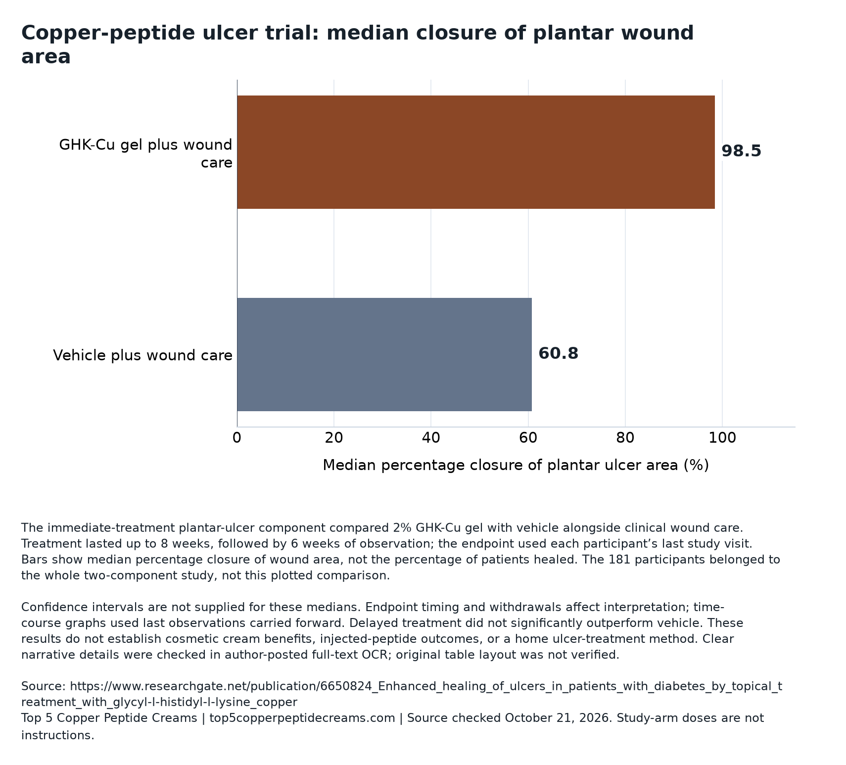
- Headline endpoint: Median wound-area closure was 98.5% with active gel versus 60.8% with vehicle in the immediate-treatment plantar component—not percentages of patients healed. [2] [1]
- Safety finding: Reported infections were 7% versus 34% in that immediate-treatment plantar subset, not across the entire trial. [2]
- Separate evidence streams: The laser study assessed postprocedure skin, while the laboratory papers examined cultured fibroblasts; neither directly tests routine facial use of a retail cream. [3] [4] [5]
What the controlled comparison actually isolated
The multicenter trial was randomized, evaluator-blinded, and vehicle-controlled. Participants received standardized debridement, pressure-relieving footwear, and education. Immediate treatment compared 2% topical gel with vehicle for up to eight weeks, followed by six weeks of observation. A delayed component used a four-week vehicle lead-in before eight additional weeks of vehicle, 2%, or 4% gel. These concentrations identify historical study arms, not instructions.
A vehicle is the formulation without the investigational active ingredient. Comparing active gel with its vehicle helps separate an ingredient-associated effect from effects of the base and the surrounding care program. The question is therefore incremental: what did adding this preparation contribute under these conditions? It is not a comparison between copper gel and doing nothing.
Interpretation also depends on what the design holds constant. If both groups receive a substantial care intervention, improvement in either group cannot automatically be credited to the investigational ingredient. Conversely, improvement in the comparison group does not invalidate an additional treatment effect. The guide to reading copper studies explains why identifying the actual comparator should precede judging the size of any result.
Who entered the trial—and who the result represents
Eligibility included adequately controlled diabetes and a full-thickness ulcer below the knee; exclusions included bone infection, gangrene, and inadequate pedal circulation. Approximately 80% of ulcers were plantar, meaning on the sole. This selected population matters: the study was not a survey of every kind of diabetic wound or every degree of vascular compromise.
A whole-study enrollment figure answers how many people entered the research program. It does not necessarily answer how many contributed to a particular comparison. Once investigators distinguish treatment timing, wound location, and starting size, a headline result may describe only one branch of the design. Reading the largest number beside the most favorable outcome can silently create a much broader claim than the analysis supports.
There is a scientific trade-off here. Restricting comparisons to more similar wounds can make an effect easier to interpret, because dissimilar causes and healing trajectories are less mixed together. But the resulting estimate becomes less representative of the full clinical spectrum. The appropriate conclusion must retain both features: better-defined relevance within a selected group and uncertainty outside it. Subgroup precision also depends on its own sample size, not on the enrollment total printed elsewhere in the paper.
Reading percentage closure without counting imaginary healed patients
Percentage area closure expresses the reduction from a wound’s starting area: subtract the remaining area from the initial area, divide by the initial area, and multiply by 100. It is a continuous measurement, capable of recording partial improvement. A healed-person proportion is a different endpoint, requiring each participant to meet a specified criterion.
Illustrative calculation—not patient data: a wound beginning at 200 square millimeters and ending at 20 has 90% area closure. It remains open. Another beginning at 40 and ending at four also has 90% closure, despite having a much smaller residual opening. The same relative result can therefore describe different absolute wound burdens.
A median adds another distinction. It identifies the middle of the ordered individual results rather than the fraction of people meeting a healing threshold. Subtracting the two reported medians gives a 37.7-percentage-point descriptive gap. That is not a patient-level treatment effect for everyone, a relative risk reduction, or a basis for calculating a number needed to treat. The table separates the main measurements so that their units do not get exchanged mid-argument.
| Measurement | Question answered | Interpretive boundary |
|---|---|---|
| Median percentage area closure | What is the middle relative area-change result? | Does not count fully healed patients |
| Categorical closure threshold | How many wounds meet a specified cutoff? | Near-complete closure need not establish durable complete healing |
| Linear edge-advance rate | How quickly does the wound margin advance after normalization? | A rate ratio is not a ratio of patients healed |
Why larger wounds and negative closure need careful arithmetic
For larger plantar ulcers, the abstract reports median closure of 89.2% versus −10.3%, with P < .01, using a size threshold above 100 square millimeters. The full-text transcription instead describes the threshold inclusively and gives vehicle expansion differing by 0.1 point. That small unresolved discrepancy should remain visible rather than be silently harmonized.
Negative closure is mathematically straightforward: the wound became larger than its baseline area. Illustratively, an opening growing from 100 to 120 square millimeters has −20% closure. This is not a negative percentage of people healing. It also reveals an asymmetry in the scale: improvement stops at complete closure, whereas enlargement has no equivalent lower bound.
That asymmetry helps explain why a mean and a median can differ substantially without either being an arithmetic mistake. A few enlarging wounds can pull a mean downward strongly; the median responds to rank order instead. Neither summary shows the entire distribution. Scientifically useful follow-up would examine individual trajectories, residual wound sizes, and uncertainty around subgroup differences. A larger apparent separation in one size category alone does not establish a biological reason that starting size modifies treatment response.
Treatment timing and closure speed answer separate questions
The delayed arms did not show significant improvement over vehicle. In the immediate plantar comparison, the report described a more-than-threefold mean closure-rate advantage using normalized linear edge advance in millimeters per day. Those are distinct findings: one concerns when treatment entered the protocol; the other concerns a geometric rate measure.
A threefold rate does not mean three times as many people healed, nor does it necessarily mean every wound required one-third as long to close. Wounds differ in shape, size, interruptions, and progression. A normalized edge measure attempts to make changing boundaries more comparable, but it remains an analytical representation rather than a direct count of completed recoveries.
Timing comparisons also raise a design problem. A lead-in period can change the population of wounds still open and the phase of recovery being observed. Early and later trajectories need not be interchangeable baselines. The observed difference therefore does not, by itself, identify a molecular explanation. Establishing why timing mattered would require evidence that distinguishes competing pathways, not simply a plausible story about tissue access or post-debridement biology. The historical result is a finding about this treatment sequence, not a general rule for applying copper products.
The endpoint clock did not stop at the same visit for everyone
The end-of-study endpoint used each participant’s last visit; longitudinal plots carried the last observation forward. This distinction separates a planned study calendar from the actual measurement used in analysis. A final recorded result is not necessarily an assessment obtained after the same elapsed time for every participant.
Carrying a last observation forward keeps an earlier measurement in later summaries. Its convenience is clear: the plotted dataset does not simply lose a person when observations end. Its assumption is stronger than it may appear, however. The method effectively treats the recorded status as a stand-in for unobserved later status. That can misrepresent subsequent improvement, deterioration, or reopening.
The direction of distortion cannot be inferred merely from knowing that the method was used. It depends on why observations ended, what happened afterward, and whether those patterns differed between groups. An illustrative concern is that departure after apparent success and departure after worsening represent very different missing trajectories. A more informative assessment would compare results under alternative assumptions about missing outcomes. Including everyone analytically is valuable, but it does not manufacture measurements that were never collected or make unequal observation times identical.
A lower infection result is not a blanket safety verdict
The report found no significant difference in overall adverse-event rates. It also specified withdrawal when an infection remained unresponsive after a week of treatment. These details belong beside the favorable subset infection result, rather than being replaced by an unqualified description of the gel as safe.
An infection endpoint and an all-event safety endpoint have different denominators and meanings. Fewer wound infections in one comparison do not establish fewer events of every kind. Nor does a nonsignificant overall comparison demonstrate identical safety: detecting uncommon harms requires sufficient exposure and reliable event ascertainment. Exact event counts and confidence intervals would help readers judge how much uncertainty remains.
Withdrawal rules additionally connect safety management to outcome interpretation. Removing someone from continued study observation may be clinically appropriate, while still leaving later wound status unknown to the analysis. This is a reason to examine the handling of follow-up, not to assume misconduct or dismiss the finding. The practical boundary is equally important: a controlled research protocol is not a home wound-care recipe. Cosmetic product documentation and general ingredient discussions cannot substitute for condition-specific clinical evaluation.
Human skin and cell studies test different propositions
The randomized laser study reported 13 completers, with enrollment and arm sizes unreported in its abstract. At 12 weeks, objective between-group wrinkle and overall-skin-quality comparisons were nonsignificant; erythema resolution also showed no significant advantage. A patient questionnaire favored GHK-Cu for perceived skin quality, P = .04. Adverse-event counts were not reported in that abstract.
That combination is not contradictory. A patient-reported endpoint measures experience; an evaluator or image-based endpoint measures a different construct. Neither should be substituted for the other. Improvement over time after a shared procedure also differs from evidence that the added regimen outperformed its comparator. The separate laser-trial analysis develops that distinction.
The culture papers add biological questions rather than settle clinical ones. One reported stimulated collagen synthesis; the other reported changes involving MMP-2 and its inhibitors. Together they suggest that matrix biology includes both production and remodeling. Moving from a cultured cell response to a visible skin benefit requires additional links: delivery to the relevant tissue, an appropriate biological response, and a meaningful clinical endpoint. A result at one level cannot simply fill every missing link.
| Evidence setting | What it investigates | What remains outside its direct test |
|---|---|---|
| Diabetic-ulcer trial | Topical gel added to standardized wound care | Routine facial cosmetic performance |
| Postlaser trial | Regimens with or without GHK-Cu after resurfacing | Everyday use on intact skin |
| Collagen fibroblast culture | Collagen synthesis in cultured cells | Visible improvement in people |
| MMP fibroblast culture | Remodeling enzymes and inhibitors | Net clinical benefit or formulation safety |
Why ingredient percentages do not produce a product ranking
The trial preparation used an HPMC gel vehicle. A shared ingredient name does not make another formulation an interchangeable version of that intervention. The relevant scientific object is the delivered preparation in its tested setting, not an ingredient detached from vehicle, route, tissue condition, and surrounding care.
This is an evidence-matching problem rather than an argument that other products cannot work. A cream might differ from a gel in ways that matter, or in ways that do not; the ulcer comparison does not determine which. Likewise, a label percentage cannot independently establish tissue exposure or comparative effectiveness. The guides to copper-peptide labels and cream, gel, or balm formats help organize those separate questions.
Editorial interpretation: product comparisons should distinguish documented terms from inferred outcomes. Readers checking brand documentation can consult the NEOVA review or CoreAge Rx review for that purpose, without treating either as evidence of ulcer treatment or trial-equivalent performance. A scientifically defensible ranking would require relevant comparative outcomes, not a chain of associations from cells to wounds to a retail jar. The same boundary prevents extrapolation from topical gel to oral or injected preparations.
What would make the evidence more decisive
Confidence intervals were not provided for the central estimates discussed here. P-values identify statistical compatibility under a testing model; they do not supply the missing range of plausible effect sizes. A result may meet a significance threshold while remaining too imprecise to support fine comparisons, especially across selected wound categories.
A more decisive evaluation would prespecify the clinically important endpoint and timing, report transparent participant flow, and distinguish complete closure from area reduction. It would also document durability, missing observations, and safety with usable denominators. These are proposed evidentiary improvements, not claims that the historical investigators collected or omitted every such measure. Original table images would resolve transcription problems but would not, on their own, answer new clinical questions.
Commercial context also deserves precision. ProCyte affiliations, the identified product, and acknowledgment of its development team document involvement; they do not establish a particular funding arrangement. Editorial interpretation: neither that relationship nor an attractive numerical result should replace appraisal of design and reporting. For purchase decisions, the cost guide can organize spending questions, but the ulcer evidence supplies no price premium justified by proven cosmetic superiority. Evidence quality is better assessed question by question than converted into a universal score.
What this evidence supports
The ulcer trial supports a treatment-associated closure signal within a particular immediate-treatment plantar comparison. Its strongest interpretive lesson is that measurement units, subgroup boundaries, and endpoint timing carry substantive scientific meaning. The evidence does not warrant treating area closure as a healed-patient rate, generalizing a subset infection finding to every participant, or ranking cosmetic products by association. The laboratory and laser studies contribute different kinds of information, not interchangeable confirmation. A useful evidence assessment preserves the positive finding while keeping its clinical question intact.
Research access and limitations
The ulcer analysis uses an indexed abstract and author-posted primary full-text OCR, not a verified original table-and-figure layout. Ambiguous arm counts and categorical proportions are therefore not reproduced. The laser and cell papers are abstract-only here; full protocols and supplements were not examined. Exact adverse-event counts were not reliably verified from the accessible ulcer transcription. Confidence intervals for the cited central estimates and a formal funding statement were not available in the reviewed material.
Your questions, answered
Does the median closure result show that almost everyone healed?
No. A median area-change measurement describes the middle individual result, not the percentage of participants with complete healing. A categorical healing analysis requires its own definition and denominator. The distinction remains important even when the median is close to the upper limit of the area-closure scale. [2]
Can the study establish lasting recovery?
Area reduction at a recorded endpoint is not automatically evidence of sustained closure afterward. Durability requires observations specifically addressing whether closure persisted. Interpretation should match the available endpoint rather than assume that a favorable final measurement settles every later outcome. [1]
Does a nonsignificant delayed-treatment comparison mean wounds never improved?
No. A nonsignificant between-group comparison concerns evidence for a difference between treatments, not whether either group changed over time. Improvement within a group and superiority over a comparator are separate questions; neither can be inferred merely from the other. [1]
Do collagen findings justify choosing the strongest copper product?
No. A culture response does not establish a useful dose-response relationship for finished products in people. Choosing a higher label percentage would require assumptions about delivery, formulation, tissue response, and clinical benefit that these laboratory experiments do not test. [4] [5]
What should readers look for in a claim citing this research?
Check whether the claim preserves the studied population, topical formulation, comparator, endpoint, and treatment context. A citation may accurately identify a real paper while still supporting a narrower proposition than the surrounding claim. No evidence here establishes that a particular marketer actually cites this trial. [2] [1]
Sources & further reading
Provider pages establish what a brand markets. Clinical and regulatory sources establish the limits of the evidence. Each source has its own last-checked date below.
- Mulder et al., 1994: GHK-Cu in diabetic neuropathic ulcers (author-posted full text)Author-posted primary full-text OCR; original table and figure layout not verified · Checked October 21, 2026
- Enhanced healing of ulcers in patients with diabetes by topical treatment with glycyl-l-histidyl-l-lysine copper.Indexed primary study abstract · Checked October 21, 2026
- Miller et al.: copper complex after CO2 laser resurfacing2006 randomized study; 13 completers; procedure-specific context · Checked October 21, 2026
- Stimulation of collagen synthesis in fibroblast cultures by the tripeptide-copper complex glycyl-L-histidyl-L-lysine-Cu2+.Indexed primary laboratory-study abstract · Checked October 21, 2026
- The tripeptide-copper complex glycyl-L-histidyl-L-lysine-Cu2+ stimulates matrix metalloproteinase-2 expression by fibroblast cultures.Indexed primary laboratory-study abstract · Checked October 21, 2026